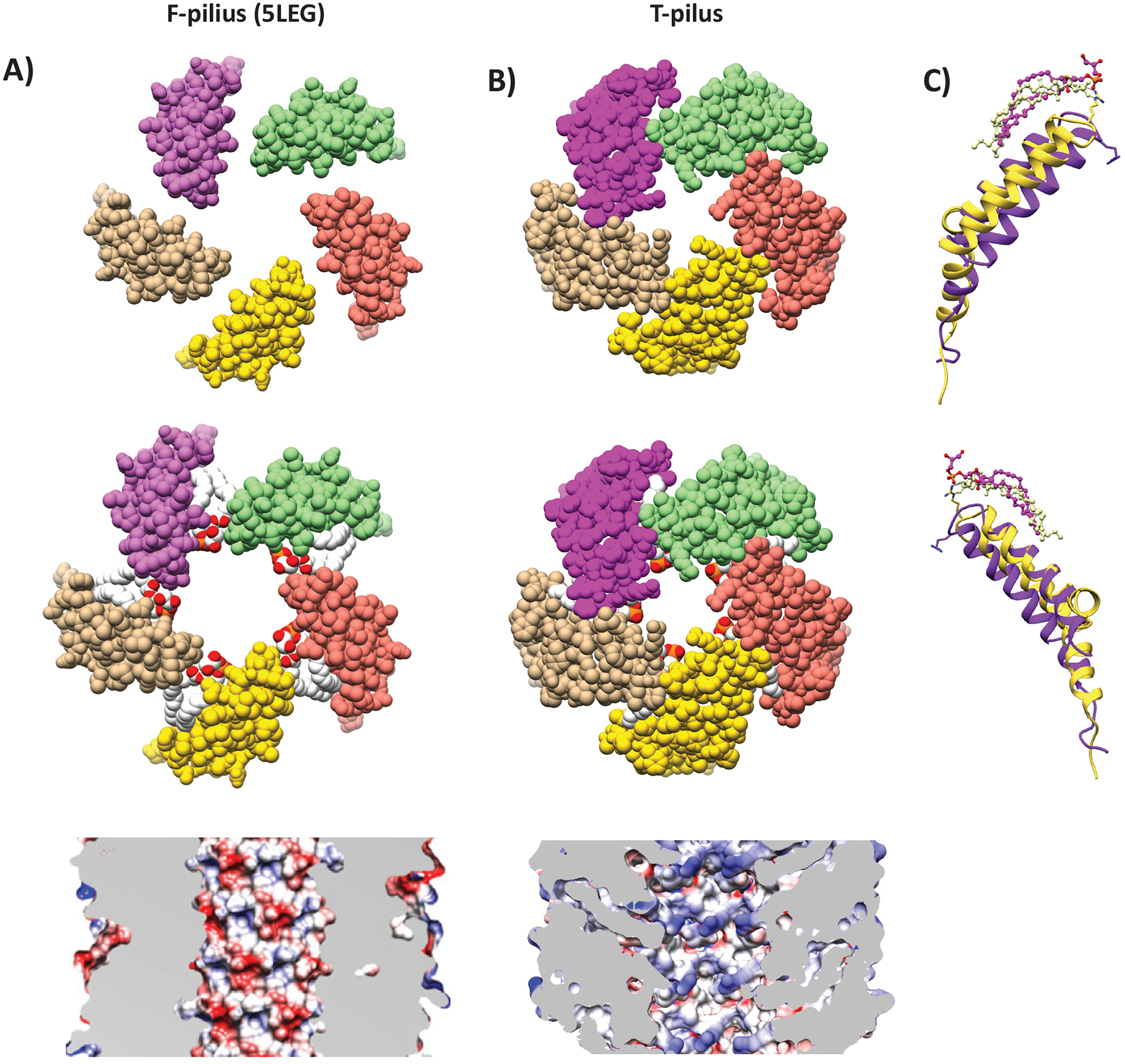
Figure 6. Structural Comparison of the T- and F-pilus.

Helical cross-sections showing organization of pilins and phospholipids in (A) F-pilus (PDB ID: 5LEG) and (B) T-pilus. The T-pilus adopts a denser structure with narrower lumen. The F-pilus lumen is moderately negatively charged (reproduced from Costa et al., 2016), whereas T-pilus lumen is neutral. (C) Structural overlay and secondary structure annotation of T-pilus VirB2 (yellow) and F-pilus TraA (purple). Phospholipids from equivalent positions in the helical packing are displayed.
