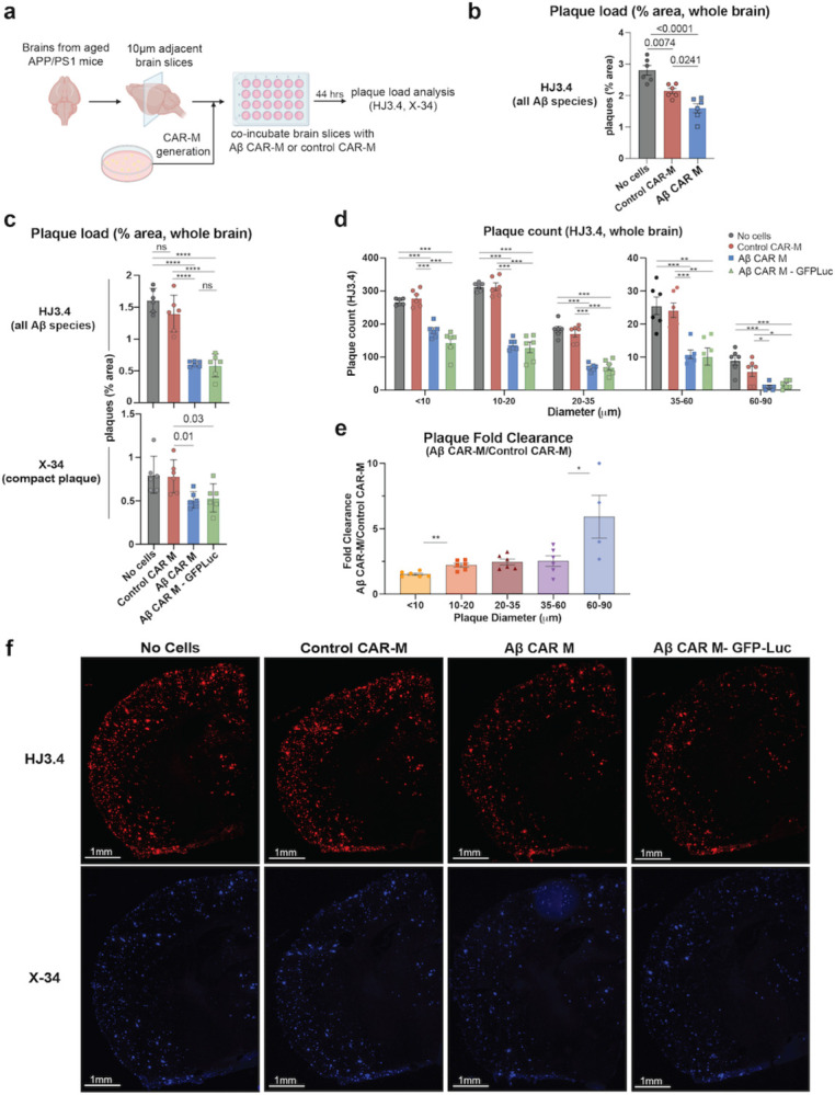
Figure 2: Aβ CAR-M resorb amyloid plaques of various sizes on brain slices from aged APP/PS1 mice ex vivo.

a) Schematic of ex vivo assessment of amyloid plaque phagocytosis. Adjacent brain slices from aged APP/PS1 mice were co-incubated with Aβ CAR-M or Control CAR-Ms for 44 hours and plaque load was assessed with HJ3.4 or X-34 immunostaining. b-e) Assessment of plaque load (b, c), plaque count (d), and plaque fold clearance of Aβ CAR-M over Control CAR-Ms (e) on APP/PS1 brain slices after co-incubation with no cells, control CAR-M, or Aβ CAR-M with or without GFP-Luc. Data shown as mean ± s.e.m from n=5–6 independent experiments with 5–6 technical replicates each. Statistical significance was calculated with one-way ANOVA with Tukey’s multiple comparisons test (c (HJ3.4), d) or unpaired t-tests (c (X-34), e). For c-e, *P < 0.05, **P < 0.01, ***P < 0.001, ****P < 0.0001. ns, not significant. f) Representative images demonstrating adjacent brain sections stained with HJ3.4 or X-34 after co-incubation with no cells, control CAR-M, Aβ CAR-M, or Aβ CAR-M GFP-Luc cells.
