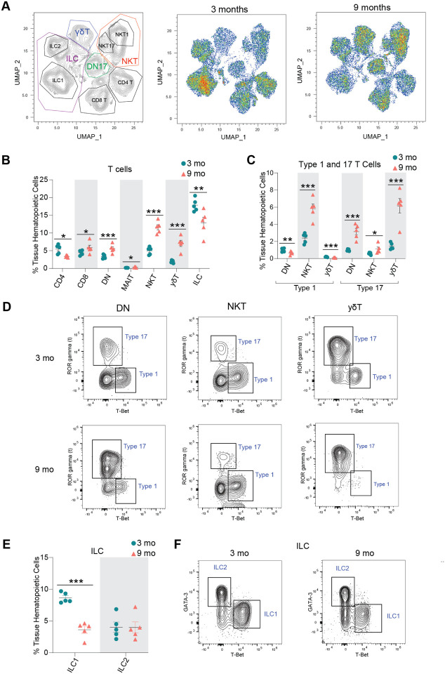
Fig. 4. Lymphocyte populations are altered in the aged ovary.
(A) UMAP of flow cytometry panel of lymphoid cells, annotated using conventional gating strategies. The number of cells plotted was normalized to better observe differences in distribution of the populations. (B) Percentage of T cell subpopulations out of tissue hematopoietic cells. (C) Percentage of Type 1 and Type 17 T cells out of tissue hematopoietic cells. (D) Representative gating of Type 1 and Type 17 lymphoid cells in young and aged ovaries. (E) Percentage of ICL1 and ILC2 out of tissue hematopoietic cells. (F) Representative gating of ILCs in young and aged ovaries. For flow cytometry, 6 ovaries from mice in the same phase of estrous cycle were pooled together to comprise each sample (n=5/age). scRNA-seq was performed in n=4 ovaries/age. Data are presented as mean ± SEM. *, **, *** represent statistical difference (FDR<0.05, 0.01 and 0.005, respectively) by multiple two-tailed t-test with Benjamini, Krieger, and Yekutieli correction for multiple comparisons.

