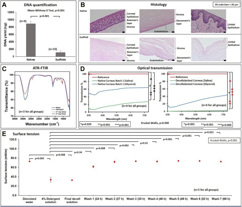
FIGURE 2.
Biochemical, microscopic, and spectroscopic assessments of native and decellularized scaffolds. (A) A graphical representation of the relative DNA contents (ng DNA/μL of homogenized tissue) in native and acellular corneas. (B) Micrographs (from a ×20 objective) illustrate the effective removal and retention of extracellular matrix compartments in decellularized corneal scaffolds compared to native tissues. (C) FTIR assessments outline the effective removal of the detergent during the washout phase from the second and third day elutes. (D) Optical transmission studies verify the ability to retain viable degrees of ocular transparency in native and decellularized corneas, compared to the reference (glass slide), which roughly supports 100% light transmission. (E) Surface tension measurements of deionized water, 4% detergent, final decell solution (collected at the end of the seventh day of the decellularization process), and washouts obtained for three consecutive days (wash 1 through wash 7).

