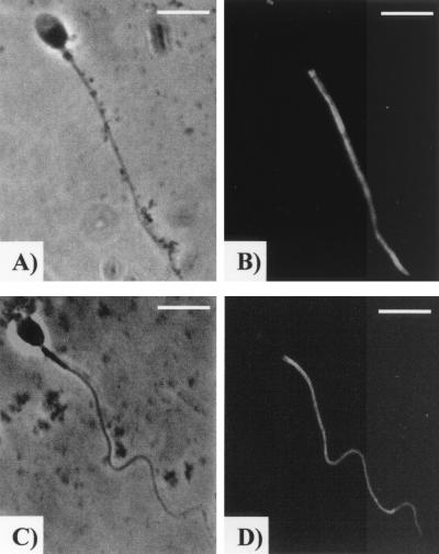
FIG. 1.
Immunolocalization of tyrosine-phosphorylated proteins on human spermatozoa. Displayed are representative examples of the major fluorescence pattern typically observed in capacitated spermatozoa (B) and spermatozoa capacitated in the presence of serovar E (D). Panels A and C are the phase-contrast images for panels B and D, respectively. Bar = 5 μm.

