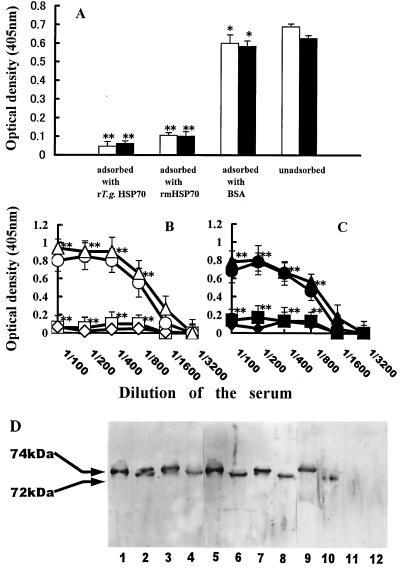
FIG. 3.
(A) Anti-mHSP70 autoantibody of T. gondii-infected mice recognized cross-reactive antigenic determinants shared by rTgHSP70 and rmHSP70. Sera of T. gondii-infected B6 mice were adsorbed with either rTgHSP70, rmHSP70, or BSA on ice as described in Materials and Methods, and then reactivities of the unadsorbed or preadsorbed sera with rTgHSP70 (open column) or rmHSP70 (closed column) were tested by ELISA. ∗, P > 0.05; ∗∗, P < 0.01 compared with the unadsorbed group. (B and C) Titration analysis of the unadsorbed and adsorbed sera of T. gondii-infected BALB/c and B6 mice with rTgHSP70, rmHSP70, or BSA against rTgHSP70 and rmHSP70. The diluted sera of T. gondii-infected mice were adsorbed with either rTgHSP70, rmHSP70, or BSA on ice as described in Materials and Methods. Titration of the sera unadsorbed (▵ or ▴) and adsorbed with either rTgHSP70 (□ or ■), rmHSP70 (◊ or ⧫), or BSA (○ or ●) against rTgHSP70 (B) and rmHSP70 (C) was analyzed by ELISA. ∗∗, P < 0.01. (D) Western blotting analysis of anti-mHSP70 autoantibody in T. gondii-infected mice. rTgHSP70 (lanes 1, 5, and 9) and TgHSP70 of Fukaya tachyzoite lysates (lanes 2, 6, and 10), rmHSP70 (lanes 3, 7, and 11), and mHSP70 of murine lymphoma line RMA lysates (lanes 4, 8, and 12) were separated by SDS-PAGE and then transferred onto nitrocellulose membranes. The membranes were probed with the sera of T. gondii-infected B6 mice (lanes 1 to 4), cross-reactive anti-TgHSP70 MAb TgCR 18 (lanes 5 to 8), and non-cross-reactive anti-TgHSP70 MAb TgNCR A5 (lanes 9 to 12).

