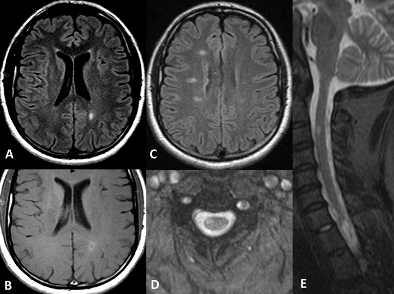
Figure 1.

A focal subcortical hyperintense FLAIR lesion ( A ) with contrast enhancement ( B ) is observed in conjunction with other periventricular and pericalosal bright lesions ( C ), similar to Dawson's fingers described for MS disease. Cervical lesions follow the same pattern, eccentrically located in the T2* axial plane ( D ) and extending for one vertebral body dimension on the sagittal STIR cervical image ( E ).
