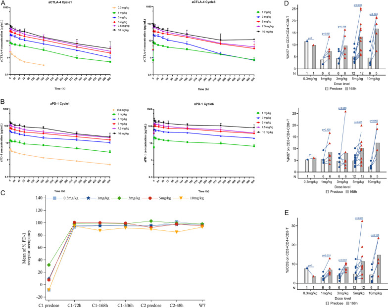
Fig. 4.
Mean (± standard deviation) plasma concentrations of anti-CTLA-4 (A) and anti-PD-1 (B) as a function of time following dosing in cycle 1 and at steady state (cycle 6) shown on a log10 scale in μg/mL across dose levels from 0.3 mg/kg to 10.0 mg/kg Q3W. C Mean % PD-1 receptor occupancy. Expression changes of Ki67 (D) and ICOS (E) on T cells in each dose group (Note, the values out of the visit window range or deviated from the protocol were not included in the summary analysis. When more than half (> 50%) of the values at a single time point are below the quantization limit (BQL), the mean values are reported as 0. For those BQL values, they are omitted on the semi-log scale plot. When there are only two samples at a single time point, the error bars are not presented

