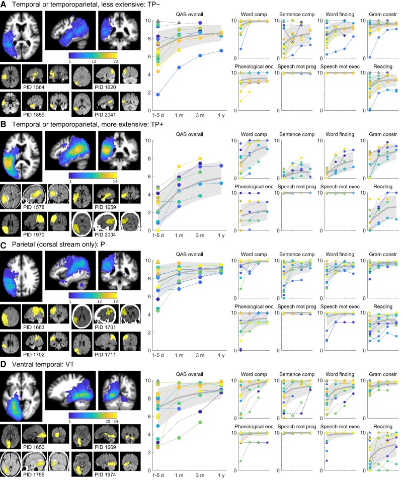
Figure 3.
Trajectories of recovery for patients with temporal and/or parietal lobe damage. (A) TP− group patients (n = 22). One patient with confirmed right hemisphere language, and no aphasia, is shown with a grey marker and does not contribute to the mean or variance estimates. (B) TP+ group patients (n = 14). (C) P group patients (n = 18). (D) VT group patients (n = 24). See Fig. 2 legend for additional details.

