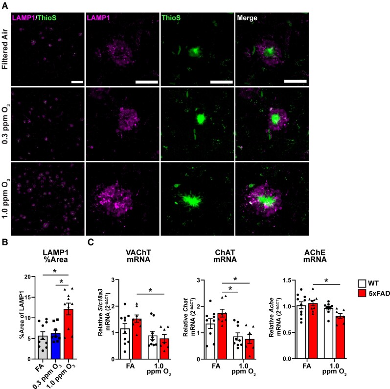
Figure 3.
Ozone exposure exacerbates neuritic dystrophy and dysregulates acetylcholinergic gene expression. 5xFAD mice (10–11 weeks old) were exposed to O3 (0.3 or 1.0 ppm) or FA by inhalation for 13 weeks. (A) Representative confocal images of ThioS (green) and LAMP1 (magenta) taken at ×20 (top) or ×63 (bottom) in the M1/M2 cortex of 5xFAD mice exposed to O3. Scale bars: top = 100 µm; bottom = 25 µm. (B) Quantification of LAMP1 per cent area. (C) mRNA expression of Slc18a3 (VAChT), Chat and Ache in the cortex of O3 exposed 5xFAD and WT mice. n = 8–10 mice/exposure group. *P < 0.05; one-way ANOVA, Bonferroni post hoc or Welch’s t-test.

