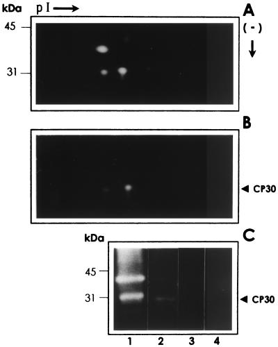
FIG. 2.
2-D substrate SDS-PAGE analyses of proteinases with affinity to HeLa cell surfaces. The proteinase patterns corresponding to the trichomonad lysates (A) and with affinity to HeLa cell surfaces obtained after a ligand assay (B) (described in Materials and Methods) were analyzed by 2-D substrate gelatin gel electrophoresis. (C) 1-D substrate gelatin gels (experimental controls) of the trichomonad lysate (2 × 10⁵ parasites) (lane 1); proteinases obtained after a ligand assay performed with the lysate from 2 × 10⁷ parasites that interacted with 1 × 10⁶ HeLa cell surfaces (lane 2); 1 × 10⁶ fixed HeLa cells used in the ligand assays that had not been exposed to parasite lysates (lane 3); and 20 μl of fresh culture medium (TYM) with horse serum (lane 4). Positions of the molecular size markers are on the left. Only the region from 50 to 25 kDa is shown. pI →, direction of isoelectrofocusing using ampholines 3/10 and 5/8 (Bio-Rad); (−) ↓, direction of the SDS-denaturing gel electrophoresis by size.

