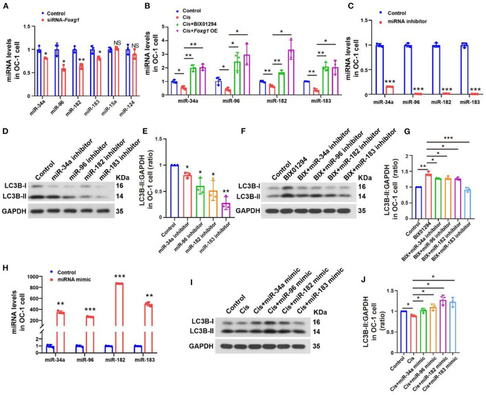
Figure 7.
Autophagy levels of OC-1 cells change through the regulation of miR-34a, miR-96, miR-182, and miR-183 expression. (A) rt-PCR of changes in miR-34a, miR-96, miR-182, and miR-183 expression with Foxg1 knockdown (B) rt-PCR of the expression in miR-34a, miR-96, miR-182, and miR-183 changes after cisplatin treatment with BIX01294 pre-treatment or Foxg1 overexpression. (C) rt-PCR of miR-34a, miR-96, miR-182, and miR-183 expression inhibition efficiency. (D) Western blotting of changes in LC3B levels after miR-34a, miR-96, miR-182, and miR-183 inhibition. (E) Quantification of the western blotting in (F). (F) Western blotting of changes in LC3B levels after BIX01294 treatment with miR-34a, miR-96, miR-182, and miR-183 inhibition. (G) Quantification of the western blotting in (F). (H) rt-PCR of miRNA mimic efficiency for enhancing miR-34a, miR-96, miR-182, and miR-183 expression. (I) Western blotting of changes in LC3B levels after cisplatin treatment with miR-34a, miR-96, miR-182, and miR-183 mimics. (J) Quantification of the western blotting in (F). *p < 0.05, **p < 0.01, ***p < 0.001.

