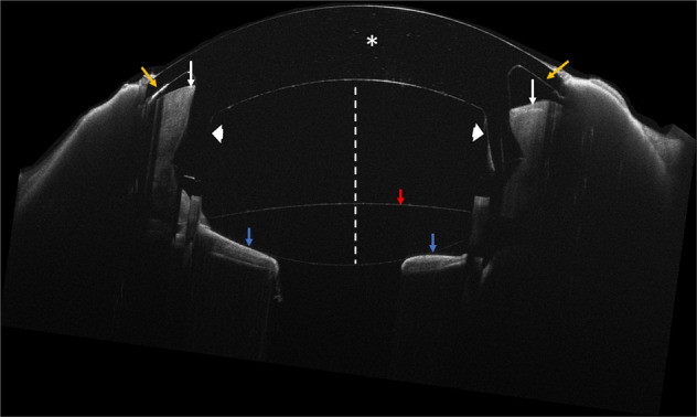
Fig. 3. Anterior segment optical coherence tomography demonstrating proper location of the CorNeat KPro at the 3-months postoperative visit.

White asterisk indicates the centrally placed optical component. White arrows indicate a cross section of the corneal remnant seating securely in the dedicated undercut of the KPro (white arrowheads). Yellow arrows indicate the integrating skirt component located underneath the conjunctiva. Note the anterior chamber (white dotted line), anterior chamber intraocular lens (red arrow) and iris (blue arrows).
