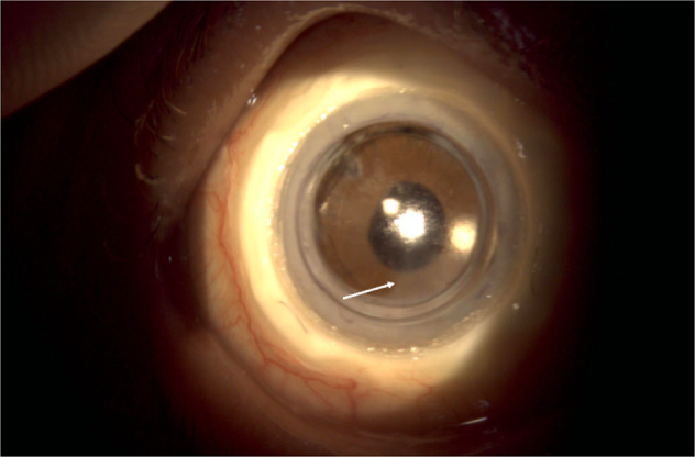
Fig. 5. An ocular photograph of the patient 6 month after the CorNeat KPro implantation.

A fine membrane over the inferior part of the anterior chamber intraocular lens (white arrow) was obsereved.

A fine membrane over the inferior part of the anterior chamber intraocular lens (white arrow) was obsereved.