Fig. 4.

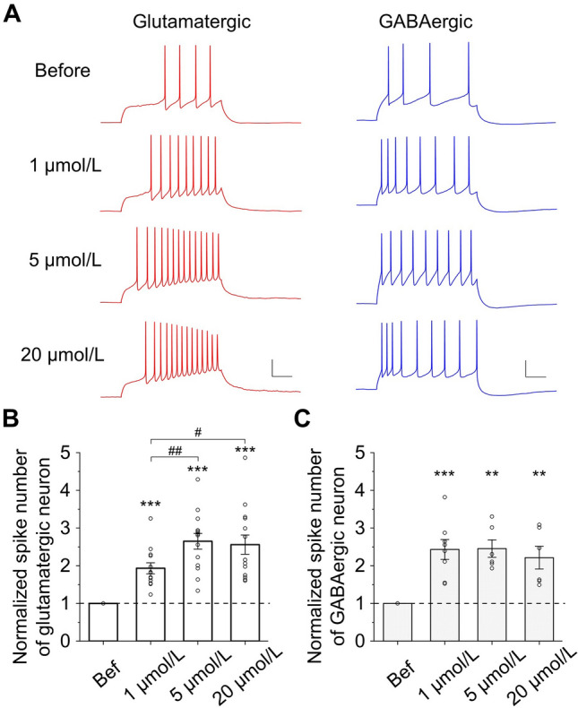
ACh increases neuronal excitability. A Example traces of action potentials induced by a depolarizing current injection (500 ms) into a glutamatergic neuron (red) and a GABAergic neuron (blue) under 4 different conditions: before, 1 μmol/L, 5 μmol/L, and 20 μmol/L ACh. Scale bars, 20 mV, 100 ms. B ACh increases the number of spikes in glutamatergic neurons (normalized to the value before ACh application. 1 μmol/L: 1.93 ± 0.15, P <0.001, n = 13; 5 μmol/L: 2.65 ± 0.21, P <0.001, n = 14; 20 μmol/L: 2.56 ± 0.25, P <0.001, n = 14; ACh vs before, paired t-test. 1 μmol/L vs 5 μmol/L: P <0.01; 1 μmol/L vs 20 μmol/L: P <0.05; 5 μmol/L vs 20 μmol/L: P = 0.79; unpaired t-test). C ACh increases the normalized number of spikes in GABAergic neurons (1 μmol/L: 2.43 ± 0.26, P <0.001, n = 8; 5 μmol/L: 2.45 ± 0.23, P <0.01, n = 6; 20 μmol/L: 2.21 ± 0.30, P <0.01, n = 6; ACh vs before, paired t-test. 1 μmol/L vs 5 μmol/L: P = 0.95; 1 μmol/L vs 20 μmol/L: P = 0.60; 5 μmol/L vs 20 μmol/L: P = 0.54; unpaired t-test).
