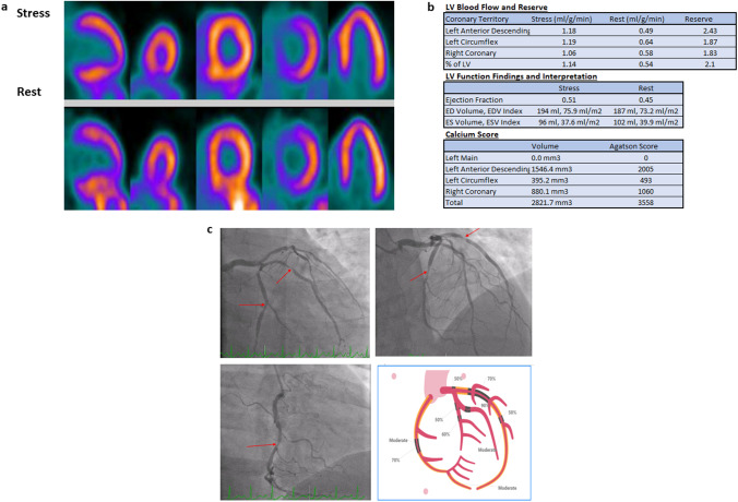
Fig. 1.
A 53-year-old male with hypertension and dyslipidemia presented with chest pain with exertion. PET relative perfusion imaging showed no defect. a The ejection fraction was 45% at rest and increased to 51% at stress. However, the total coronary artery calcium scoring was 3558, with calcifications noted in the LAD, LCx, and RCA. The global myocardial flow reserve was borderline (2.1), primarily because of reduced stress flow (1.14 ml/g/min). b Balanced ischemia was suspected, and the patient was referred for Invasive angiography, which showed multivessel disease with obstructive stenosis in the distal LM, proximal-to-mid LAD, proximal-to-mid LCx, and mid-RCA (red arrows). The patient subsequently underwent triple vessel CABG (LIMA to LAD, SVG to PDA, and SVG to OM). c On a recent follow-up visit 1.5 years from CABG, the patient continued his guideline-directed therapy, experienced no incident cardiovascular events, and reported no symptoms

