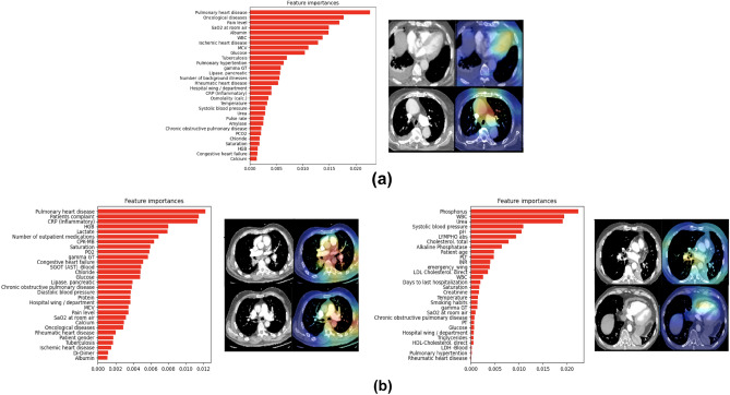
Figure 5.
Misclassified examples: (a) False negative example. (b) Two false positive examples. For each example, the EHR selected features from the multimodal classifier arrange by importance gain are presented on the left. On the right—representative Grad-CAM visualizations. (a) The heatmaps overlay the heart area, specifically the LV and also the pulmonary artery (PA). The EHR features indicate a number of illnesses that support this such as pulmonary hypertension and other heart conditions. (b) The false positive examples indicate acute PE and overall severe illness as well focusing on the pulmonary truck, Aorta (left), RV chamber and also covering the PE clot (right) with EHR feature supporting this.

