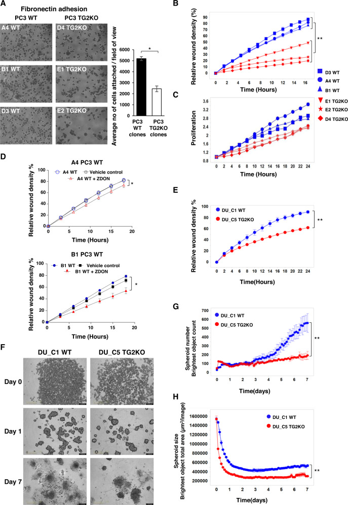
Fig. 2. TG2KO decreases cell adhesion, migration and spheroid formation of metastatic PC3 and DU145 cells.
A Cell attachment for 1 h on fibronectin of WT and TG2KO PC3 clones evaluated by manual counting of the number of attached cells from three fields of view (FOV) per clone. Data represent the mean of three independent experiments performed in quadruplicates. B Cell migration assay of three PC3 WT and three TG2KO clones grown to confluence (40,000 cells/well) in ImageLock 96-well plates and wound created using WoundMaker®. Time course of wound closure is expressed as relative wound density (%), which is a measure of cell density in the wound area relative to the density of cells outside of the wound area. Data are representative of three independent experiments performed in quadruplicates. C PC3 WT and TG2KO clones were grown on standard 96-well plates to assess proliferation. Time lapse course of proliferation is expressed as phase area confluence. Data normalised to the start of experiment, 0 h, p ≥ 0.18 (ns). D Cell migration assay of PC3 WT (A4 and B1 clones) treated with irreversible TG2 inhibitor ZDON and vehicle control (DMSO), to assess the effect on migration. The wound healing assay was performed as described in (B). E Cell migration assay of DU145 WT and TG2KO clones (DU_C1 WT and DU_C5 TG2KO) performed as described in (B). F Spheroid formation assay representative images of same DU145 WT and TG2KO clones. Cells were seeded in ultra-low attachment plate at 4000 cells/well, centrifuged and spheroid formation monitored using the IncuCyte® S3 live cell imaging from day 0 to day 7. G Reduced number of spheroids and H decreased size of spheroids formed by DU_C5 TG2KO compared to DU_C1 WT clone. Error bars represent ± SEM. Student’s t-test: *p ≤ 0.05; **p ≤ 0.01.

