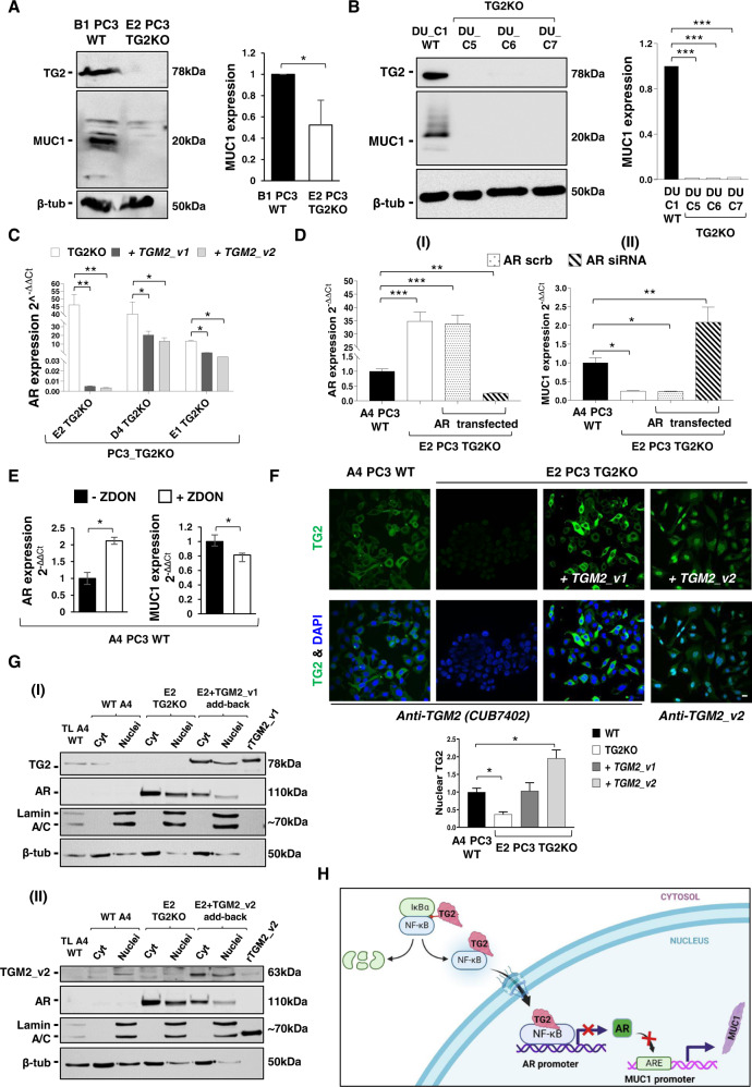
Fig. 4. Modulation of MUC1 expression by TG2 via the androgen receptor in metastatic PCa cell lines.
A, B Immunoblot showing decreased protein expression of MUC1 in TG2KO clones from metastatic PCa cell lines, PC3 and DU145. Densitometry analysis of MUC1 expression normalised versus β-tubulin loading control and WT cells, Student’s t-test: *p ≤ 0.05, ***p ≤ 0.001. C Relative quantifications of Androgen Receptor (AR) in three representative PC3 TG2KO clones (D4 PC3 TG2KO, E1 PC3 TGKO and E2 PC3 TG2KO), transfected back with either TGM2_v1 or TGM2_v2 in each clone. Values obtained from relative quantification of AR were normalised to the expression of AR in a WT clone (A4 PC3 WT). D Effect of AR knockdown on MUC1 expression. Relative expression of AR (I) and MUC1 (II) following siRNA knockdown of AR 48 h post transfection in E2 PC3 TG2KO clone compared to A4 PC3 WT. House-keeping gene, HPRT. E AR and MUC1 expression following 24 h pharmacological inhibition of TG2 using irreversible TG2 inhibitor ZDON (90 µM) in A4 PC3 WT clone. All qRT-PCR were analysed using Student’s t-test: *p ≤ 0.05, **p ≤ 0.01. F Nuclear localisation of TGM2_v2 protein in E2 PC3 TG2KO clone after being transiently transfected, as shown by immunofluorescence. E2 PC3 TG2KO cells transfected with pcDNA3.1(+)ValTGM2_v1 and pcDNA3.1/Hygro(-)TGM2_v2 for 48 h via electroporation. Cells were washed and fixed with ice-cold 90% methanol and incubated with primary antibody (either mouse monoclonal CUB7402, 1:200 or custom-made rabbit polyclonal anti-TGM2_v2, 1:200) for 16 h, followed by incubation with respective secondary antibodies conjugated with FITC, 1:200. Cells were then visualised using Leica SP5 confocal microscope. Quantification of nuclear staining using Image J Fiji software, Student’s t-test: *p ≤ 0.05. G Cytosolic and nuclear fractions of A4 PC3 WT clone, E2 PC3 TG2KO clone either transfected or not with TGM2_v1 (I) or TGM2_v2 (II) cDNAs were fractionated using the NE-PER nuclear and cytoplasmic extraction reagents kit (Thermo Scientific). Equal amounts (35 µg) of A4 WT total cell lysate (TL) and fractions were separated in a 8% SDS-polyacrylamide gel under reducing conditions and membranes probed with anti-TG2 CUB7402 (I) or anti-TGM2_v2 antibody (II) and AR. Loading controls were lamin A/C for the nuclear fractions and β-tubulin for the cytosolic fractions. TL: total cell lysate. H Schematic depiction of the mechanism of MUC1 regulated expression via TG2 and AR expression in PCa cells. In PCa cells TG2 leads to IĸBα degradation with release of its subunits (p65/p50) [27]. The TG2/p65/50 complex translocates into the nucleus [28] and inhibits AR expression: without AR the mucin-1 promoter is not blocked and this leads to MUC1 transcription. This schematic was created with BioRender.

