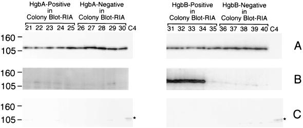
FIG. 8.
Western blot-based detection of the HgbA, HgbB, and HgbC proteins in hemoglobin-grown isolates of N182. Equivalent amounts of whole-cell lysates from the 20 BHI-Hg-grown isolates were probed with HgbA-specific MAb 17H3 (A); HgbB-specific MAb 4B3 (B); and HgbC-reactive MAb 12A2 (C). The BHI-Hg-grown isolates identified in the colony blot RIA as positive for HgbA expression are in lanes 21 to 25. The BHI-Hg-grown isolates that were negative for HgbA expression in the colony blot RIA are in lanes 26 to 30. The BHI-Hg-grown isolates positive for HgbB expression in the colony blot RIA are in lanes 31 to 35. The BHI-Hg-grown isolates that were negative for HgbB expression in the colony blot RIA are in lanes 36 to 40. Control lane C4 in panel C contains a lysate from N182 cells grown in BHI to allow detection of HgbC. The asterisk beside lane C4 denotes the location of the HgbC protein. It should be noted that the HgbC-reactive MAb bound to a doublet in this Western blot; the upper band of the doublet (marked with the asterisk) is the HgbC protein. Molecular size markers (in kilodaltons) are on the left.

