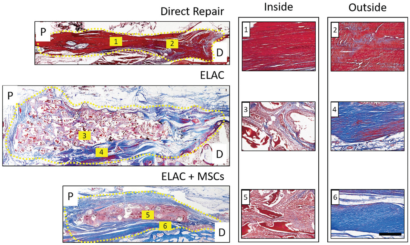
Figure 4.

Representative histological sections of the direct repair, ELAC, and ELAC + MSC groups stained with Masson trichrome (blue depicts de novo and loosely packed collagen; red highlights ELAC and other extracellular matrix components). Proximal (P) and distal (D) regions are noted, and the region of the repair is highlighted by a dotted yellow line. Collagen was more difficult to distinguish in the direct repair specimens, but it was still visible (blue and red regions overlap in 1 and 2). Collagen within the continuum of the scaffolds for both scaffold groups (faint blue visible in 3 and 5) was not as robust or apparent as collagen in the tissue surrounding the scaffold structure (deep blue present in 4 and 6). Scale bar = 200 mm. ELAC, electrochemically aligned collagen; MSC, mesenchymal stem cell.
