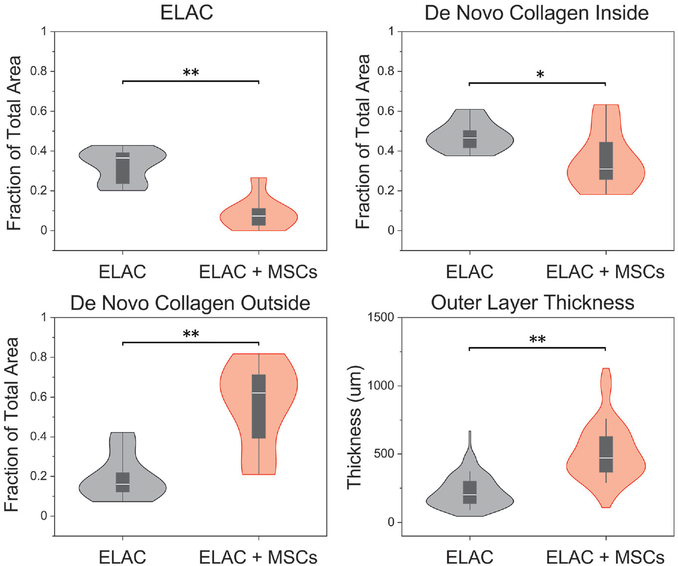
Figure 5.

Violin plots illustrating the distributions of the fraction of total area occupied by ELAC, de novo collagen inside, and de novo collagen outside, as well as measurements of the thickness of the outer layer of tissue surrounding the scaffold. Significantly more de novo collagen outside the continuum of the scaffold structure was noted for the ELAC + MSC group and less of the total area was occupied by ELAC fibers in the ELAC + MSC group. Measurements of the thickness of the layer of tissue surrounding the scaffolds were significantly higher in the ELAC + MSC group. Significance noted as (**) for P <.05 and (*) for .05 ≤ P ≤ .10. ELAC, electrochemically aligned collagen; MSC, mesenchymal stem cells.
