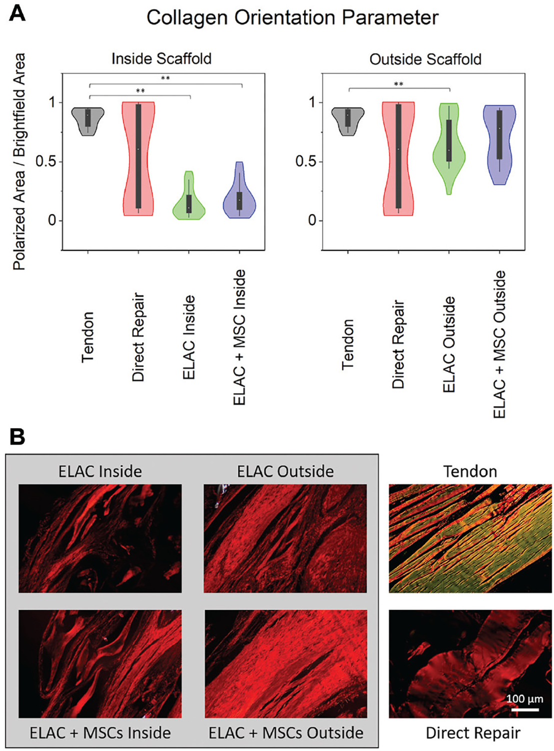
Figure 6.

(A) Violin plots highlighting the distributions for assessment of orientation of the collagen fibers in histological specimens stained with picrosirius red and imaged under polarized and brightfield conditions. On the y-axes, “0” indicates no tissue visible under polarized light, and “1” indicates that all of the tissue visible under brightfield conditions is visible under polarized light (or aligned). Collagen within the continuum of the scaffold structure showed significantly lower alignment compared with the native tendon for both scaffold groups; however, collagen outside the continuum of the scaffolds was similar to the native tendon for the ELAC + MSC group. (B) Representative polarized light microscopy images from representative specimens demonstrating the orientation of the collagen fibers. Scale bar = 100 μm. Significance noted as (**) for P <.05. ELAC, electrochemically aligned collagen; MSC, mesenchymal stem cell.
