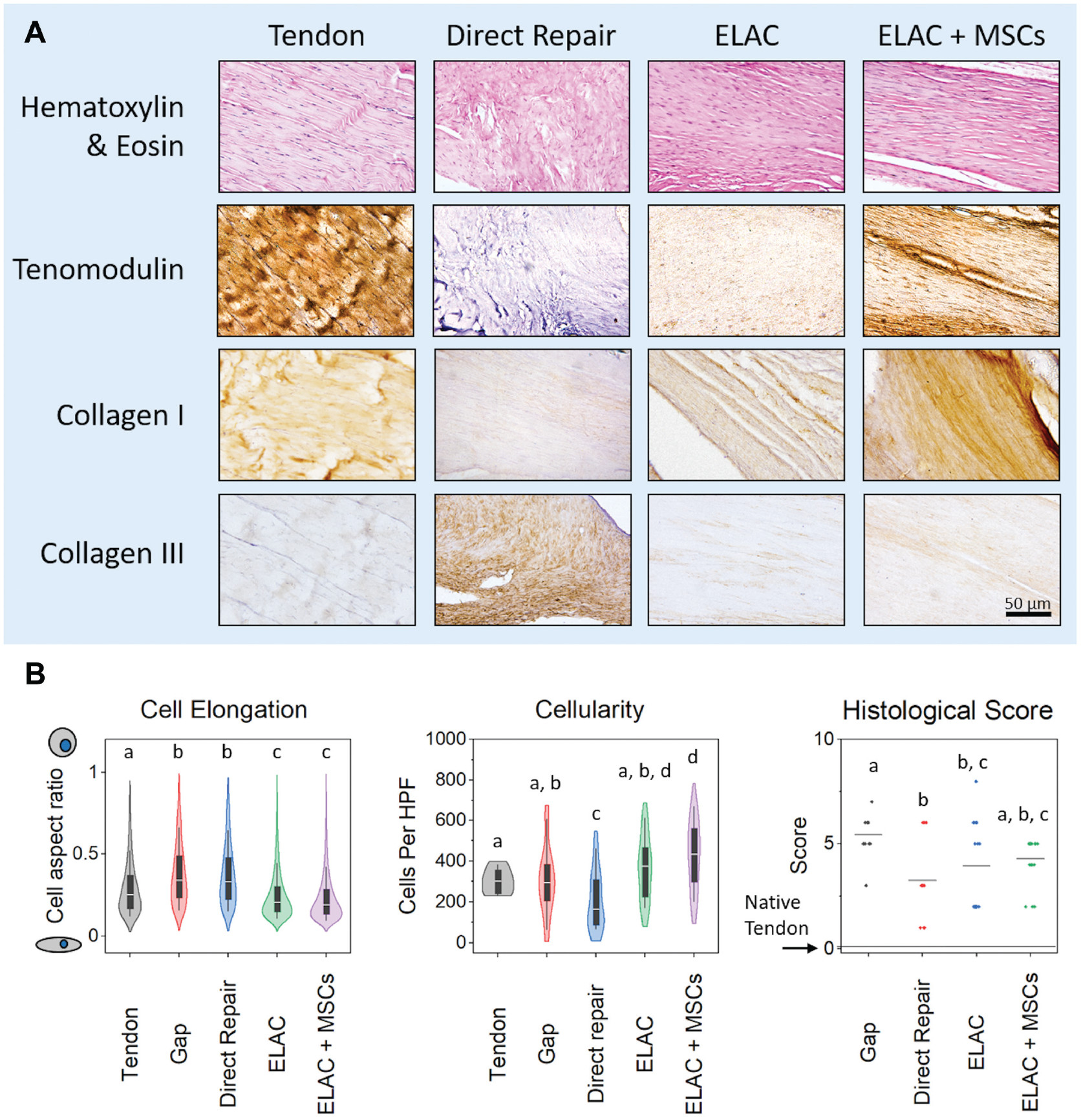
Figure 7.

(A) Histological and immunohistochemical staining. Hematoxylin and eosin staining shows the cellularity and cellular elongation/shape within the tissue surrounding the ELAC scaffolds. Tenomodulin was present in the ELAC and ELAC + MSC groups and notably absent in the direct repair group. Collagen type 1 appeared to be present in greater amounts in the scaffold-containing groups as compared with the direct repair group, and staining for collagen type 3 illustrated the opposite trend (more was present in the direct repair group than in ELAC scaffold groups). Scale bar = 50 μm. (B) Histological metrics. Significantly more cells were elongated in the ELAC and ELAC + MSC groups when compared with the native tendon, gap, and direct repair groups. In terms of cellularity, the direct repair group had the fewest cells per high-power field of view, and the ELAC + MSC group had significantly more cells than all groups except for the ELAC group. Histological scores for tendon-like metrics revealed that the gap group was significantly less tendon-like than the other 3 operative groups (a score of “0” indicates no apparent difference from the native tendon). Significance in (B) is noted by letters above each group in the plot that corresponds to the grouping. For example, a group denoted with the letter a is not statistically different from another group that is also denoted with a. ELAC, electrochemically aligned collagen; MSC, mesenchymal stem cell.
