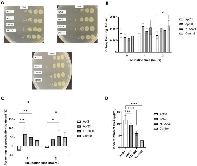
Figure 4.
Effect of aptamers on the survival of P. aeruginosa. (A) Representative images of drop plate method for analysis of antibacterial activity of aptamers on P. aeruginosa growth. Each plate represents different time points (0, 1 and 2 h) starting from the dilution 104 (right) to 109 (left). (B) Colony forming unit (CFU/mL) of P. aeruginosa in the presence and absence of 10 µM of aptamers (n = 5). (C) Percentage of P. aeruginosa growth after 1- and 2-h incubation with and without aptamers. (D) Concentration of DNA released by P. aeruginosa after 2 h incubation with 10 μM of aptamers indicating leakage of cytoplasmic materials due to membrane damage (n = 3). Data represent an average of three independent experiments, ± SD shown by error bar; *p value of < 0.05, and **p-value of < 0.01.

