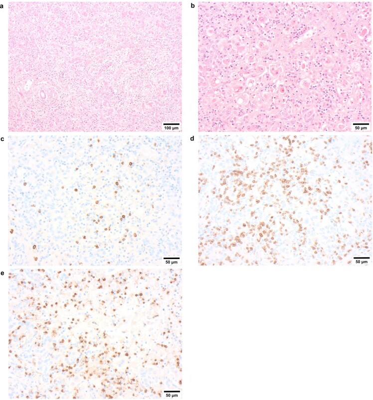
Extended Data Fig. 5. Representative histology of case livers.
a & b, H&E sections x100 and x200 showing a pattern of acute hepatitis with parenchymal disarray, there is a normal, uninflamed, portal tract lower left image a. Spotty inflammation and apoptotic bodies are shown in b along with perivenular hepatocyte loss/necrosis. Immunohistochemistry shows fewer mature B lymphocytes (CD20 panel c) than T lymphocytes (CD3, panel d, pan T cell marker) most of which are cytotoxic CD8 lymphocytes (panel e). In conclusion the livers of these children have a distinctive pattern of damage which does not indicate a specific aetiology, it does not exclude but does not offer positive support for either autoimmune hepatitis or a direct cytopathic effect of virus on hepatocytes. Each image shows a representative result from histology carried out on a minimum of five cases.

