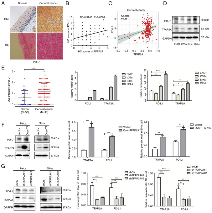
Figure 2.
TFAP2A positively regulates PD-L1 expression in cervical cancer. (A) IHC staining of PD-L1 expression and hematoxylin-eosin staining in normal and cancer tissues. (B) The association between PD-L1 and TFAP2A protein (semi-quantitative analysis using IHC staining). (C) The association between PD-L1 and TFAP2A protein in The Cancer Genome Atlas database. (D) Western blot analysis of TFAP2A and PD-L1 expression in normal (END1) and cervical epithelial (HeLa, C33a and SiHa) cell lines. (E) Reverse transcription-quantitative PCR of TFAP2A and PD-L1 expression in normal (END1) and cancer) cervical epithelial (HeLa, C33a and SiHa) cell lines. (F) Western blot analysis revealed that PD-L1 expression was reduced in TFAP2A-overexpressing cervical cancer cell lines. (G) Western blot analysis revealed that PD-L1 expression was reduced in cervical cancer cell lines subjected to TFAP2A knockdown. n=3 for cell culture analysis. *P<0.05, **P<0.01 and ***P<0.001. TFAP2A, transcription factor AP-2 alpha; PD-L1, programmed death-ligand 1; H&E, hematoxylin-eosin; IHC, immunohistochemical.

