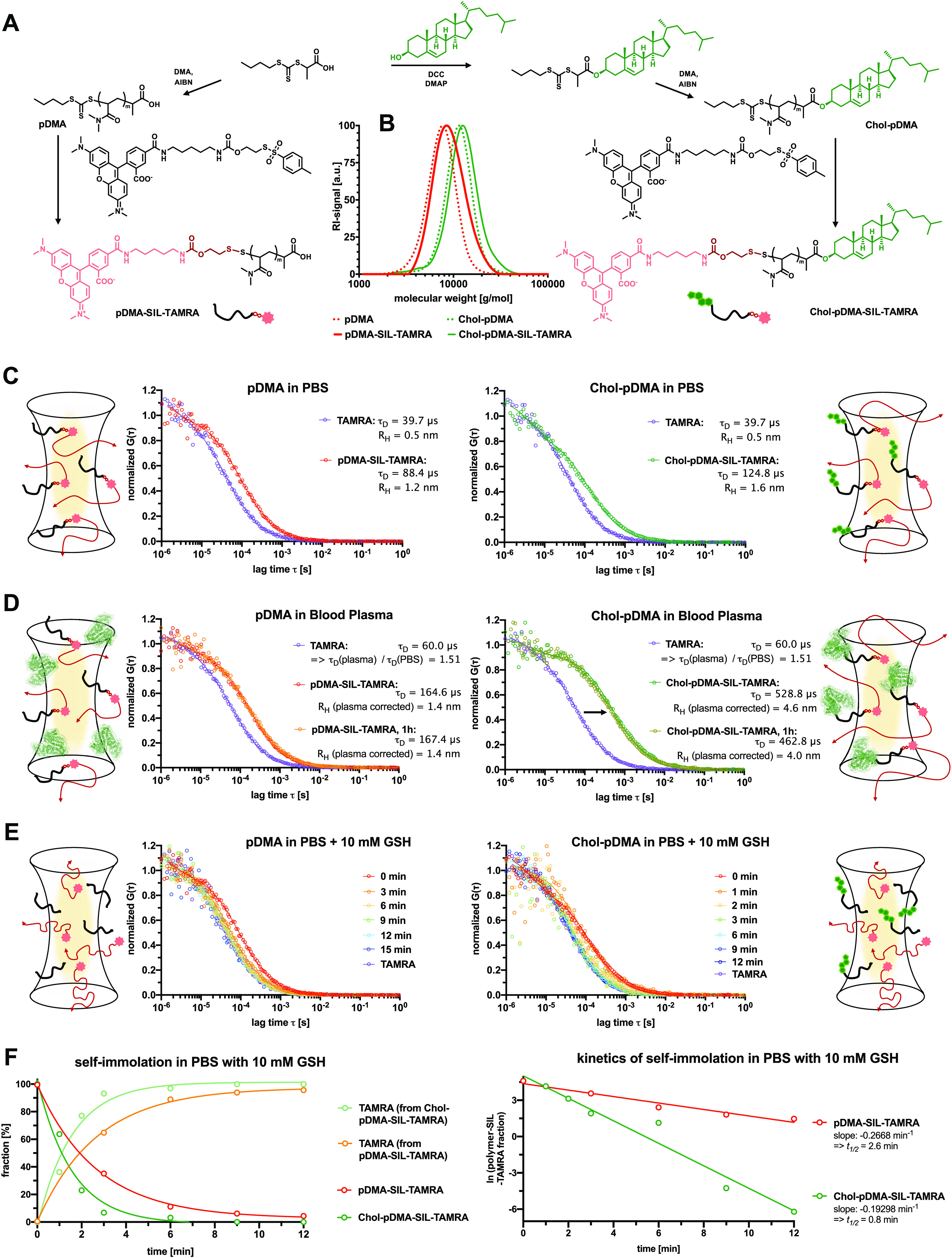
Figure 5.

Comparison of the RAFT polymer-derived SIL-TAMRA conjugates with and without α-end cholesterol modification. (A) Synthetic scheme of the chain-transfer agent modification with or without cholesterol prior to RAFT polymerization of DMA (targeting an average DP = 50). The resulting polymers with the cholesterol α-end group exhibit improved membrane permeability and capability of noncovalently binding to albumin, while the trithiocarbonate end group can still be exploited for SIL-TAMRA conjugation. (B) Successful TAMRA conjugation does not alter the polymer distribution verified by SEC. (C) Fluorescence correlation spectroscopy (FCS) measurements of pDMA-SIL-TAMRA (left) and Chol-pDMA-SIL-TARMA in PBS and their corresponding results. (D) FCS measurements of pDMA-SIL-TAMRA (left) and Chol-pDMA-SIL-TARMA in full human blood plasma. Both conjugates remain stable and do not release the dye. While pDMA-SIL-TAMRA remains as individual polymer chains in full blood plasma (left), Chol-pDMA-SIL-TARMA exhibits lager sizes indicating interactions with plasma protein components including albumin. (E) FCS measurements of pDMA-SIL-TAMRA (left) and Chol-pDMA-SIL-TARMA in PBS with 10 mM glutathione (GSH) indicating the rapid reductive-responsive release of the dye. (F) A fit of two fluorescent species was applied to these measurements for quantifying the fraction of polymer–TAMRA conjugates and released TAMRA (left). These data can further be analyzed by first-order release kinetics (right) revealing a half-life of about t1/2 = 2.6 min for pDMA-SIL-TAMRA and t1/2 = 0.8 min for Chol-pDMA-SIL-TAMRA.
