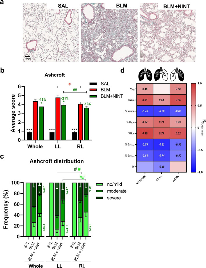
Fig. 5.
Histological assessment of fibrosis progression in BLM model and Nintedanib effect. A Representative images of Masson’s trichrome stained histological sections from SAL (left), BLM (middle), and BLM + NINT (right) mice at 21 days. B Ashcroft score (AS) quantification for the SAL, BLM, and BLM + NINT groups in the whole, left (LL), and right (RL) lungs as indicated. Statistical significance of the differences between groups for the whole lung were calculated by Two-way ANOVA followed by Dunnett’s t post-hoc test (*p < 0.05; ***p < 0.001 vs. BLM group), whereas a Student’s t-test analysis was used to evaluate the statistical significance of AS differences between the left and the right lobe of the same group (#p < 0.05; ##p < 0.01). C AS class frequency distribution (no/mild, moderate, and severe) for the whole, left, and right lungs. Statistical differences between the class frequencies of BLM and BLM + NINT groups were evaluated via Two -way ANOVA followed by Dunnett’s t post-hoc test (*p < 0.05). Student’s t-test analysis was used to evaluate the statistical significance of AS class frequency differences between the left and the right lobe of the same groups (#p < 0.05; ##p < 0.01). D Heat-map representation of Spearman correlation coefficients (R) between CT-derived parameters and AS in the whole, left, and right lungs; no significant correlations (p-value < 0.05) have not been reported

