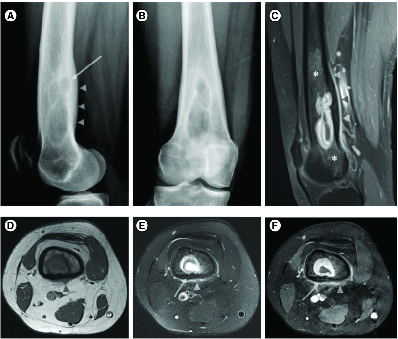
Figure 1. . X-ray of distal right femur and coronal postcontrast fat suppressed images.
X-ray of distal right femur in lateral (A) and AP (B) views showing an ill-defined expansile lytic lesion in the right distal femoral shaft with endosteal scalloping and posterior cortical sclerotic reaction and thickening (arrow and arrowhead in (A), respectively). Coronal postcontrast fat suppressed sagittal (C) and axial (F) T1 WI and axial T1 (D) and fat suppressed T2 images of the right distal femur showing a rim enhancing lesion with central fluid signal. There is cortical thickening and invasion evidenced by loss of the normal low signal in the cortical bone (arrow in (E & F)), associated with endosteal scalloping and smooth periosteal/soft tissue extension (arrow heads in (C–F)). Note the surrounding enhancing marrow signal abnormality worrisome for lesion extension more than red marrow islands (asterisks in (C)).

