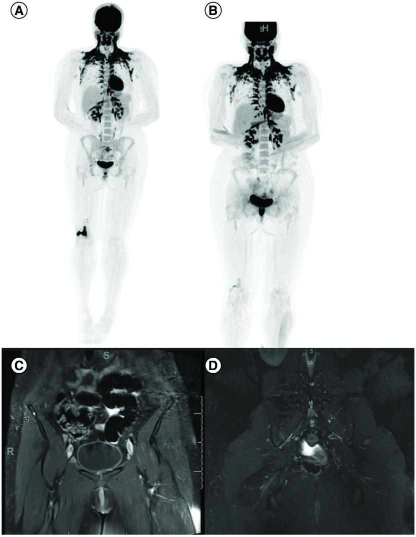
Figure 3. . Images showing multiple extra CNS lesions.
Intensely FDG-avid lytic lesion of the right distal femur (A) as well as lytic metastases involving the right popliteal region, right groin, right external and common iliac chains. Improvement in sacrum and right femoral FDG uptake after treatment (B). Note, physiological brown fat activation in the neck. Coronal contrast enhanced fat suppressed T1 WI showing corresponding punctate enhancing metastatic nodules in the right iliac bone (C) and left S1 level (D).

