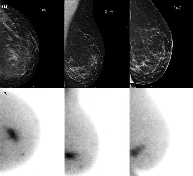
Fig. 2.
Breast imaging of a 56-year-old patient with left-sided breast cancer, detected at 1-year follow-up after treatment for right-sided breast cancer. With digital mammography, more glandular tissue was described in the medial lower quadrant (BI-RADS 3) (a), but no mass could be distinguished and ultrasound was negative. Molecular breast imaging (MBI; Dilon 6800 gamma camera) revealed suspicious sestamibi uptake in the medial lower quadrant over an area of 36 mm (b). MBI-guided biopsy was conducted, showing invasive carcinoma grade 2 with a lobular growth pattern, hormone receptor-positive, HER2-negative.

