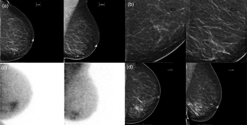
Fig. 3.
Breast imaging of a 69-year-old patient with left-sided breast cancer and ductal carcinoma in situ. The patient was referred by the national breast cancer screening program due to incomplete imaging (BI-RADS 0). There appeared to be slightly more glandular tissue in the medial lower quadrant on digital mammography (a) including the mammographic enlargements (b), with an inhomogeneous echotexture, but no mass could be distinguished. Adjunct molecular breast imaging showed suspicious irregular sestamibi uptake in the medial lower quadrant (c) (Dilon 6800 gamma camera). A hydromarker was placed in the area with inhomogeneous echotexture to confirm correlation with the pathological sestamibi uptake on MBI (d), after which ultrasound-guided biopsy was performed. The representativity of the acquired biopsy specimens was confirmed by showing uptake of sestamibi in the specimens in vitro. Pathological analysis showed ductal carcinoma in-situ grade 3 with a small focus of invasive carcinoma, hormone receptor-negative, HER2-positive. MBI, molecular breast imaging.

