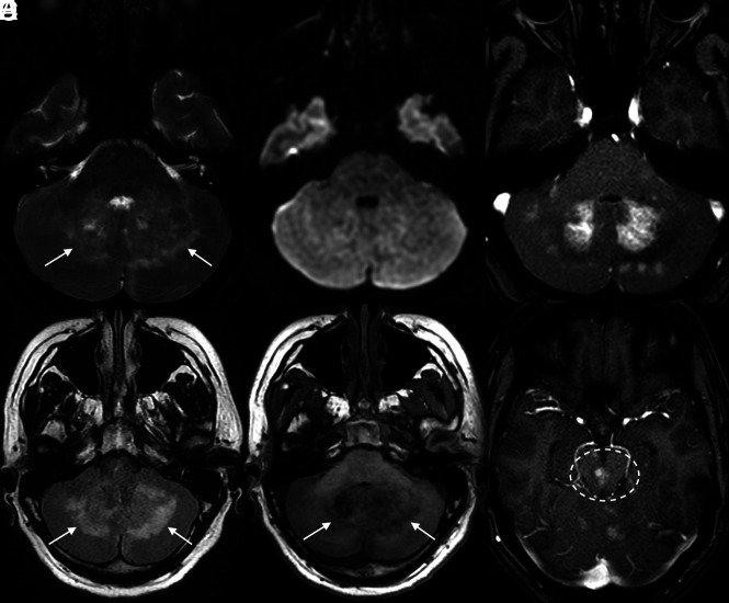
FIG 1.
MR images of the posterior fossa demonstrate multifocal abnormalities involving both cerebellar hemispheres, particularly located around the dentate nuclei. These regions are bright on T2 and FLAIR (arrows in A and B), lack restricted diffusion (C), are hypointense on T1 (arrows in D), and demonstrate patchy enhancement on postcontrast T1 fat-saturated images (E). Similar patchy enhancement is also noted in the midbrain (dashed oval on F).

