FIGURE 19.

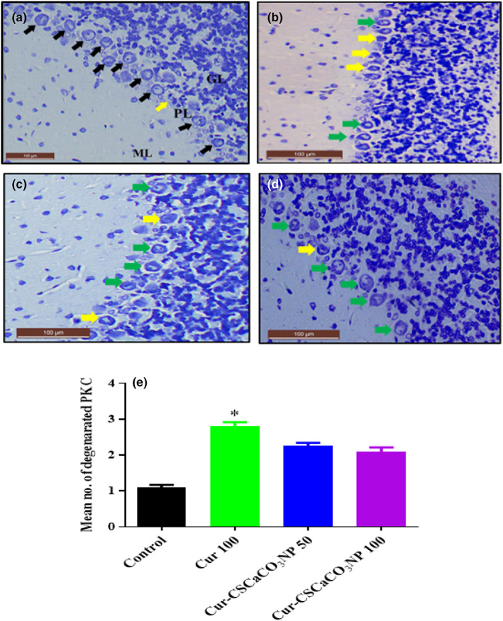
Toluidine blue‐stained cerebellum sections from rats treated with (a) normal saline (control) showing the normal layers architecture molecular layer (ML), Purkinje layer (PL), and granular layer (GL): And normal piriform‐shaped Purkinje cells (black arrow). (b) 100 mg/kg of curcumin (cur 100) showing vacuolar spaces and irregular Purkinje cells with eosinophilic cytoplasm (yellow arrow). (c) 50 mg/kg of cur‐CSCaCO3NP (cur‐CSCaCO3NP 50) showing fewer damaged Purkinje cells. (d) 100 mg/kg of cur‐CSCaCO3NP (cur‐CSCaCO3NP 100) showing marked improved layers organization and restoration of normal Purkinje cell morphology (green arrow). (e) Quantitative representation of degenerated Purkinje cells of the control and all the treated groups. *p < 0.05 versus control, n = 5. (Toluidine blue, ×20, scale bar = 100).
