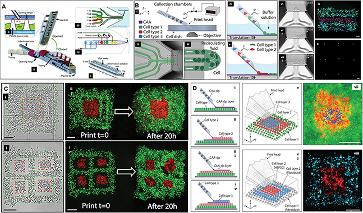
Figure 6.

A) Schematic diagram showing a (i) PDMS microfluidic pipette tip, (ii) the holder, the (iii) pneumatic interface between the holder and the tip, wells, and (iii-iv) the microfluidic switching junction near the tip. Reproduced from [206] with permission. B) (i) The microfluidic printhead is connected a controller hardware and software interface, which regulates the deposition of various cell types at a time, by allowing for the formation of a (ii-v) recirculating fluid zone at the end of the printhead within a liquid bath. (vi-viii) The translational movement of the printhead and X and Y, coordinated with the adhesion of the cells to treated substrate, allows for the patterning of cells with (ix) multi- or (x) single cell resolution. C) The bioprinter enables fabrication of multi-cellular two-dimensional (planar) tissues, in this case skin cancer cells (A431, red) surrounded by epithelial cells (HaCaT, green). The scale bars represent 200 μm. D) Three-dimensional tissue constructs where (v,vii) the base cell layer was composed of A431 cells (green), the middle layer being HaCaT cells (red), and the top layer being A431 cells (blue); in (vi,viii), a patch of liver cancer cells (HepG2, in red) surrounded by fibroblasts (3T3-J2, in blue). The scale bars represent 300 μm. Reproduced from [207] with permission.
