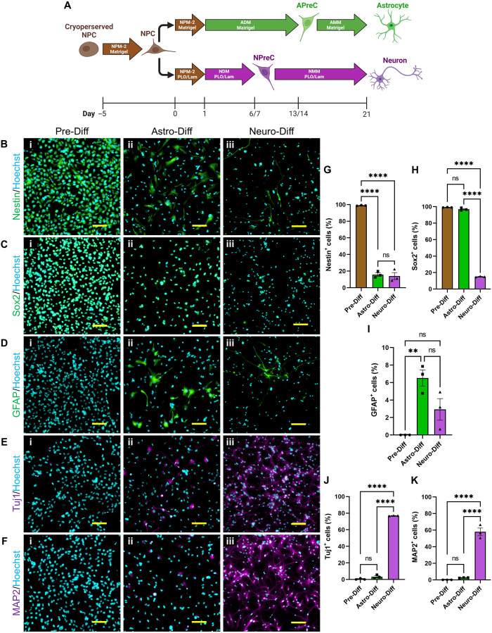
Fig. 1. NPC differentiation and characterization.
(A) Diagram of NPC differentiation in 2D cell culture. In the astrocyte differentiation protocol, astrocyte differentiation medium (ADM) was used to convert NPCs to astrocyte precursor cells (APreCs). Astrocyte maturation medium (AMM) was then used to convert APreCs to NPC-derived astrocytes (NPC-ACs). In the neuron differentiation protocol, neuron differentiation medium (NDM) was used to convert NPCs to NPreCs. Neuron maturation medium (NMM) was then used to convert NPreCs to NPC-derived neurons (NPC-neurons). Both differentiation cultures were terminated on day 21. Culture dishes were coated with Matrigel or poly-l-ornithine and laminin (PLO/Lam) where indicated. (B to F) Immunocytochemical analysis of NPC differentiation. Cells were immunofluorescently labeled for the neural stem cell markers, nestin (B, green) and Sox2 (C, green), the AC marker, GFAP (D, green), and the neuron markers, Tuj1 (E, purple) and MAP2 (F, purple) before any differentiation (Pre-Diff, i), as well as after astrocyte (Astro-Diff, ii) and neuron (Neuro-Diff, iii) differentiation protocols. Nuclei were labeled with Hoechst (blue). Scale bars, 100 μm. (G to K) Graphs showing the percentage of total cells that expressed nestin (G), Sox2 (H), GFAP (I), Tuj1 (J), and MAP2 (K) in Pre-Diff, Astro-Diff, and Neuro-Diff cultures. The data show mean value, error bars ± SEM, n = 3, one-way analysis of variance (ANOVA) with Tukey’s test; not significant (ns), P > 0.05; **P < 0.01; ****P < 0.0001.

