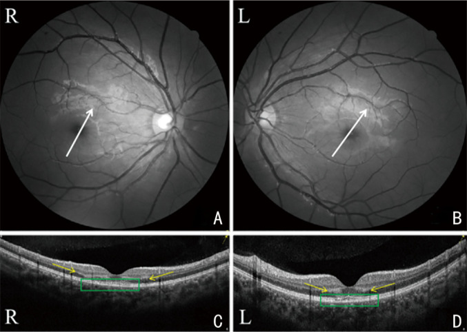
Figure 2. OCT B-scan images of case No.4, corresponding to the white arrows in fundus photography (A and B): localized outer plexiform layer and outer nuclear layer (yellow arrows) of the macula are hyper-reflective, and the lesion was more evident in the right eye (C) than left eye (D), and the elipsode zone and interdigitation zone are damaged more evident in the left eye (green rectangle).

