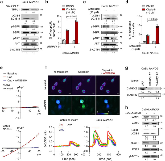
Fig. 5. TRPV1 promotes cisplatin resistance through Ca2+-AMPK pathway in NANOG+ tumor cells.
a, b CaSki NANOG cells were transfected with siRNA targeting GFP or TRPV1. c, d CaSki NANOG cells were treated with DMSO or AMG9810. a, c The levels of TRPV1, LC3B, pEGFR, EGFR, pAKT, and AKT proteins were confirmed by western blot analysis. b, d The frequency of apoptotic (active caspase 3+) cells were analyzed by flow cytometry. e Current-voltage relationships in capsaicin (Cap, red line) or AMG9810 (AMG, blue line)-induced whole-cell current in CaSki no insert versus CaSki NANOG cells. f Fluorescence calcium imaging by TRPV1 activity in tumor cells. Top, representative calcium images of FURA-2-AM-loaded cells. Bottom, changes in intracellular Ca2+ current over time in tumor cells. g, h CaSki NANOG cells were transfected with siRNA targeting GFP or CaMKKβ. The expression levels of indicated protein were confirmed by western blots. a, c, g, h β-actin was used as the internal loading control. Numbers below the blot images indicate the expression as measured by fold-change. All experiments were performed in triplicate. The p values by two-way ANOVA (b, d) are indicated. The data represent the mean ± SD. Source data are provided as a Source Data file.

