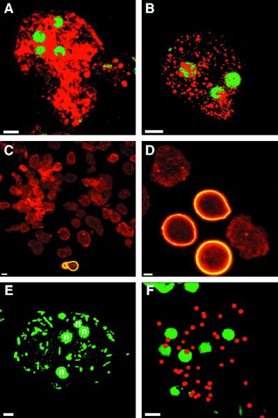
FIG. 8.
Confocal micrographs of wall-less cysts, which were made by encysting E. invadens parasites for 2 days in the presence of 50 mM galactose. Wall-less cysts, which were permeabilized before labeling, had four nuclei (stained with Sytox green in panels A, B, E, and F) and contained numerous secretory vesicles stained with anti-Jacob antibodies (red in panel A) and anti-chitinase antibodies (red in panel B). The vast majority of wall-less cysts, which were labeled with anti-Jacob antibodies (red in panel C), lacked chitin on their surface, which was detected with WGA (yellow in panel C). In contrast, many control parasites encysting in the absence of galactose were spherical and bound WGA to their cyst walls (yellow in panel D). The galactose lectin was present on the surface of wall-less cysts, which phagocytosed GFP-labeled bacteria (stained green in panel E) or mucin-coated beads (stained red in panel F) when excess galactose was removed. Bars, 5 μm.

