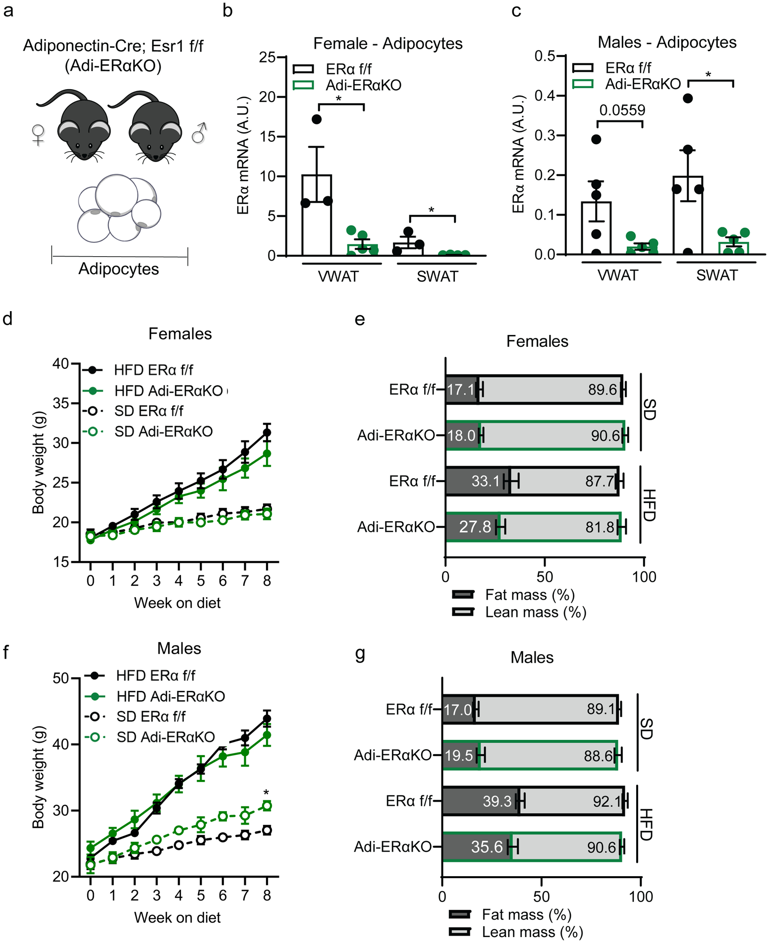
Figure 1. Adipocyte ERα deletion does not impact body composition.

(A) Experimental mouse model to delete ERα in mature adipocytes using the Adiponectin-cre; Esr1 fl/fl;mTmG strain.
(B) ERα expression in isolated mature adipocytes from Adi-ERα-KO females and controls. (n=3–5 mice per group)
(C) ERα expression in isolated mature adipocytes from Adi-ERα-KO males and controls. (n=5 mice per group)
(D) Body weight of female Adi-ERα-KO and controls during 8 weeks of SD or HFD. (n= 6–7 mice per group)
(E) Body composition of female Adi-ERα-KO and controls after 8 weeks of SD and HFD. (n=6–7 mice per group)
(F) Body weight of male Adi-ERα-KO and controls during 8 weeks of SD and HFD. Comparison shown is between SD groups. (n= 5–6 mice per group)
(G) Body composition of male Adi-ERα-KO and controls after 8 weeks of SD and HFD. (n=5–6 mice per group)
Statistical significance determined by unpaired t-tests in panel B-C and two-way ANOVA with Tukey’s test for panels C and E. *p<0.05. Error bars represent mean ± S.E.M. SD: standard diet, HFD: high-fat diet, VWAT: visceral fat, SWAT: subcutaneous fat. See also Figure S1.
