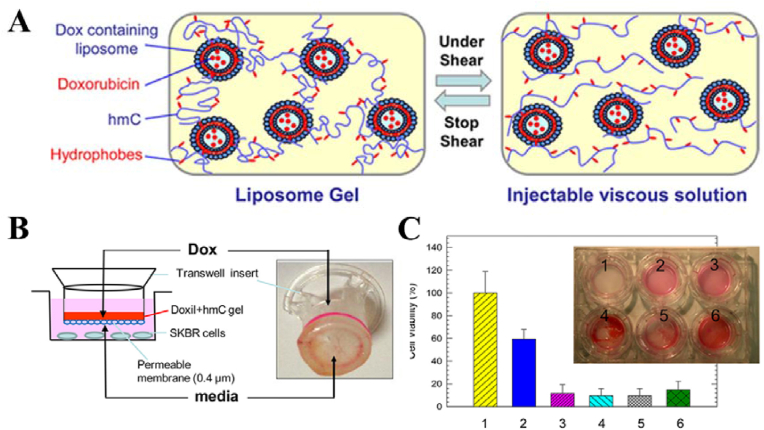
Fig. 28.
(A) Structure of an injectable liposome gel at rest (left) and under shear force (right). (B) Schematic of the experimental setup. SK-BR-3 cells are plated per well, and a given sample is added to the upper chamber. The samples are (1) DMEM media (control); (2) solution of 13 mg/mL hmC; (3) solution of hmC+ Dox (180 μg/mL); (4) gel of hmC+ Doxil (Dox, 400 μg/mL); (5) gel of hmC+ Doxil (Dox, 320 μg/mL); and (6) gel of hmC+ Doxil (Dox, 280 μg/mL). (C) Cell viability results and photographs from trypan blue staining after 11 days. Reproduced with permission [107]. Copyright 2012, American Chemical Society. (For interpretation of the references to color in this figure legend, the reader is referred to the Web version of this article.)

