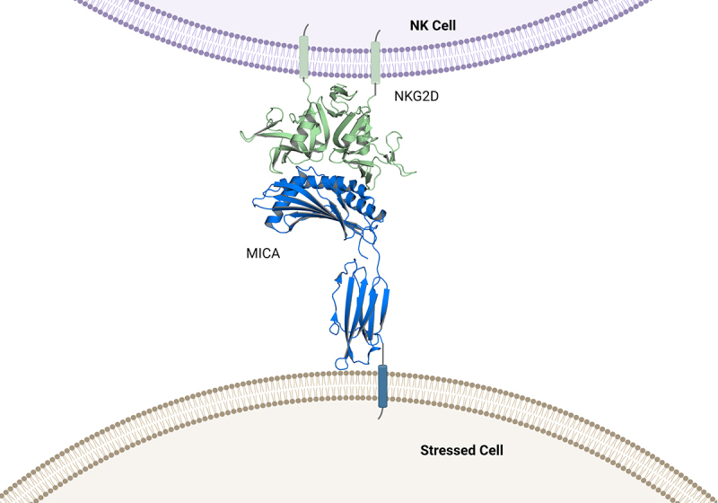
Figure 3.

Structure of the NKG2D-MICA complex. .
MICA is shown in blue. The alpha 1 and alpha 2 domains of MICA form the interface with the NKG2D receptor, depicted in green. The arrow indicates the region containing proteolytic cleavage sites of MICA which are proximal to the cell membrane. (Figure adapted from https://www.rcsb.org/, PDB 1HYR (complex of MICA and NKG2D).
