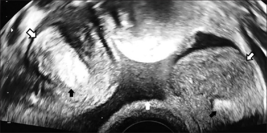
Figure 7.

Two-dimensional vaginal ultrasound, axial section at the level of the upper limit of the uterine horns, showing 2 uterine cavities (black arrows) and 2 uterine horns (open arrows pointing the serosa, white arrow pointing free fluid lying on the uterine fundus, interposed between the 2 uterine horns)
