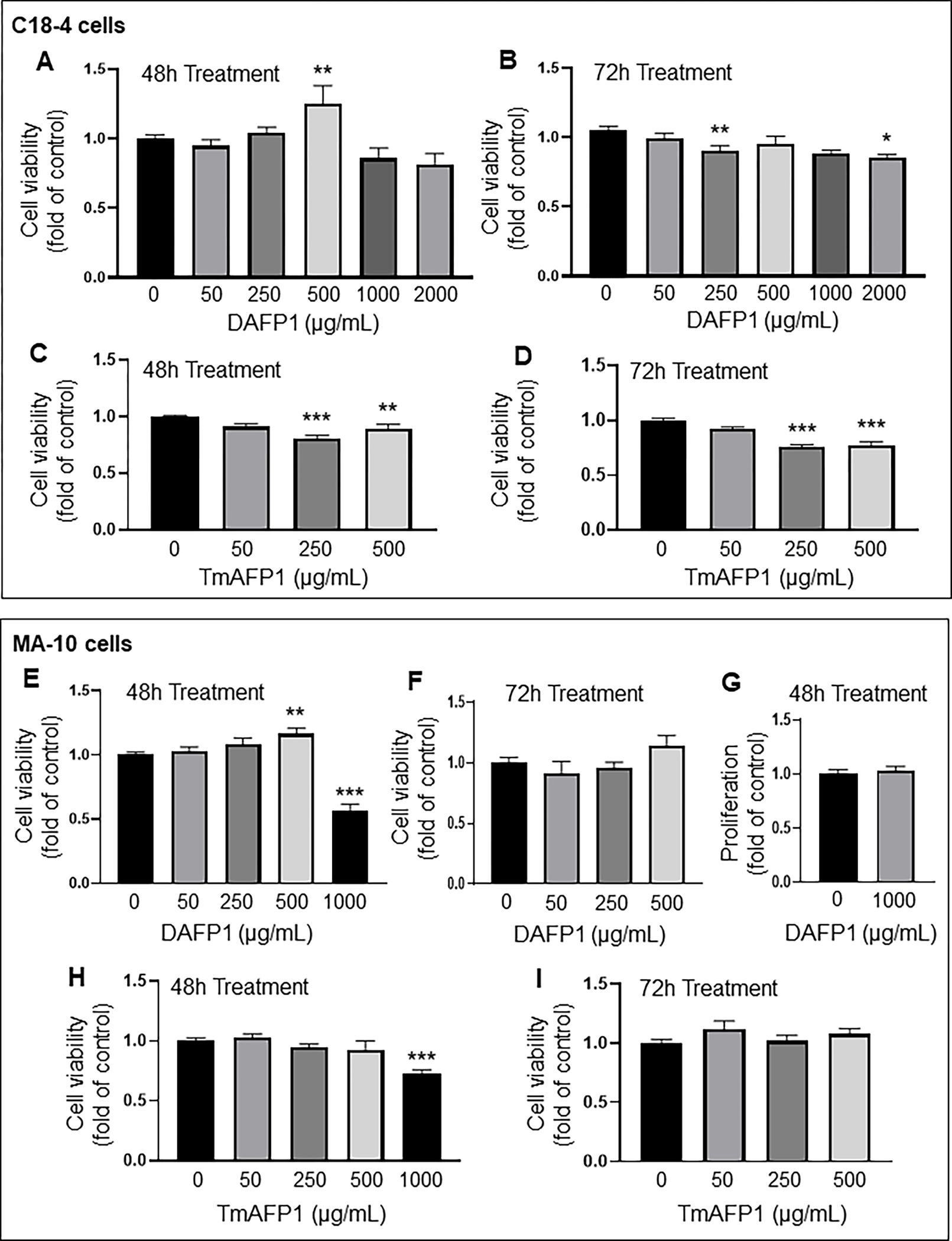
Figure 1. Effect on AFPs on the viability of testicular C18-4 spermatogonia and MA-10 Leydig cells.

(A-B) Effect of 0 to 2000 μg/mL DAFP1 on C18-4 cell viability measured by MTT assay over (A) 48h and (B) 72h treatments. (C-D) Changes in viability with TmAFP treatments at 0 to 500 μg/mL over (C) 48h and (D) 72h. (E-G) Effect of 0 to 1000 μg/mL DAFP1 on MA-10 cell viability measured by MTT assay over (E) 48h and (F) 72h treatments. (G) MA-10 cell proliferation measured by EDU incorporation assay in control cells (0) and cells treated with 1000 μg/mL DAFP1 for 48h. (H-I) Changes in MA-10 cell viability with TmAFP treatments at 0 to 1000 μg/mL over (H) 48h and (I) 72h. Results are presented as fold change of vehicle control. N=3 independent experiments conducted in triplicates. Significant difference relative to vehicle control with One way ANOVA test and multiple comparisons: * (p≤0.05), ** (p<0.01), *** (p<0.001).
