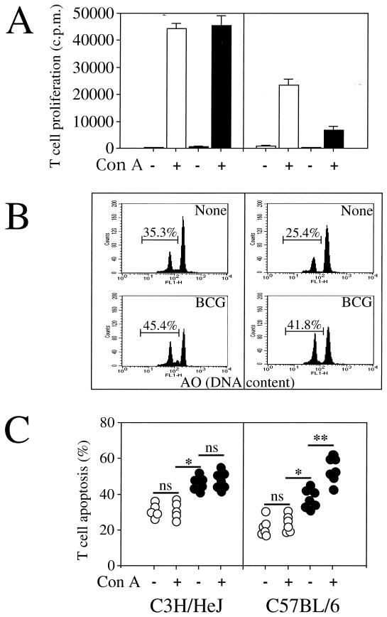
FIG. 3.
The defect of T-cell proliferation in BCG-infected C57BL/6 mice is related to T-cell apoptosis. T cells were purified from spleens of uninfected (white bars) or BCG-infected (black bars) C3H/HeJ and C57BL/6 mice 2 weeks after intravenous infection with 5 × 10⁶ CFU of BCG. The cells were cultured and restimulated in vitro in the absence (−) or presence (+) of ConA. (A) Proliferative T-cell responses were measured by [3H]thymidine incorporation. The data represent the means of values from pools of three mice assayed in triplicate. Results are from one of two independent experiments with similar results. (B) The percentage of spontaneous T-cell apoptosis from individual mice was measured by FCM analysis using acridine orange (AO) nuclear dye. T-cell apoptosis was also determined by light microscopy analysis (data not shown). Both determinations gave similar results, with less than a 5% difference between the two methods, as previously described (11). (C) T-cell apoptosis of uninfected animals is represented by the open circles, and T-cell apoptosis of BCG-infected animals is represented by the solid circles. Each symbol represents the result from an individual animal. Significant difference in spontaneous T-cell death between uninfected and infected mice is represented by ∗ (P < 0.05); significant difference in T-cell death between nonstimulated and activated T cells is represented by ∗∗ (P < 0.05). ns, not significant.

