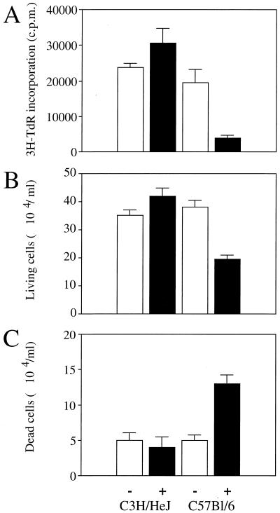
FIG. 5.
T cells from both uninfected (−) and BCG-infected (+) C3H/HeJ and C57BL/6 mice were cultured in the presence of anti-CD3 antibodies. (A) Lymphoproliferation was assessed after 3 days of activation with anti-CD3 antibodies, by measuring the incorporation of [3H]thymidine (3H-TdR). (B and C) Viable (B) and dead (C) T cells were counted under a light microscope after 72 h of in vitro activation. The data represent the means of values from pools of three animals and are representative of three independent experiments with similar results.

