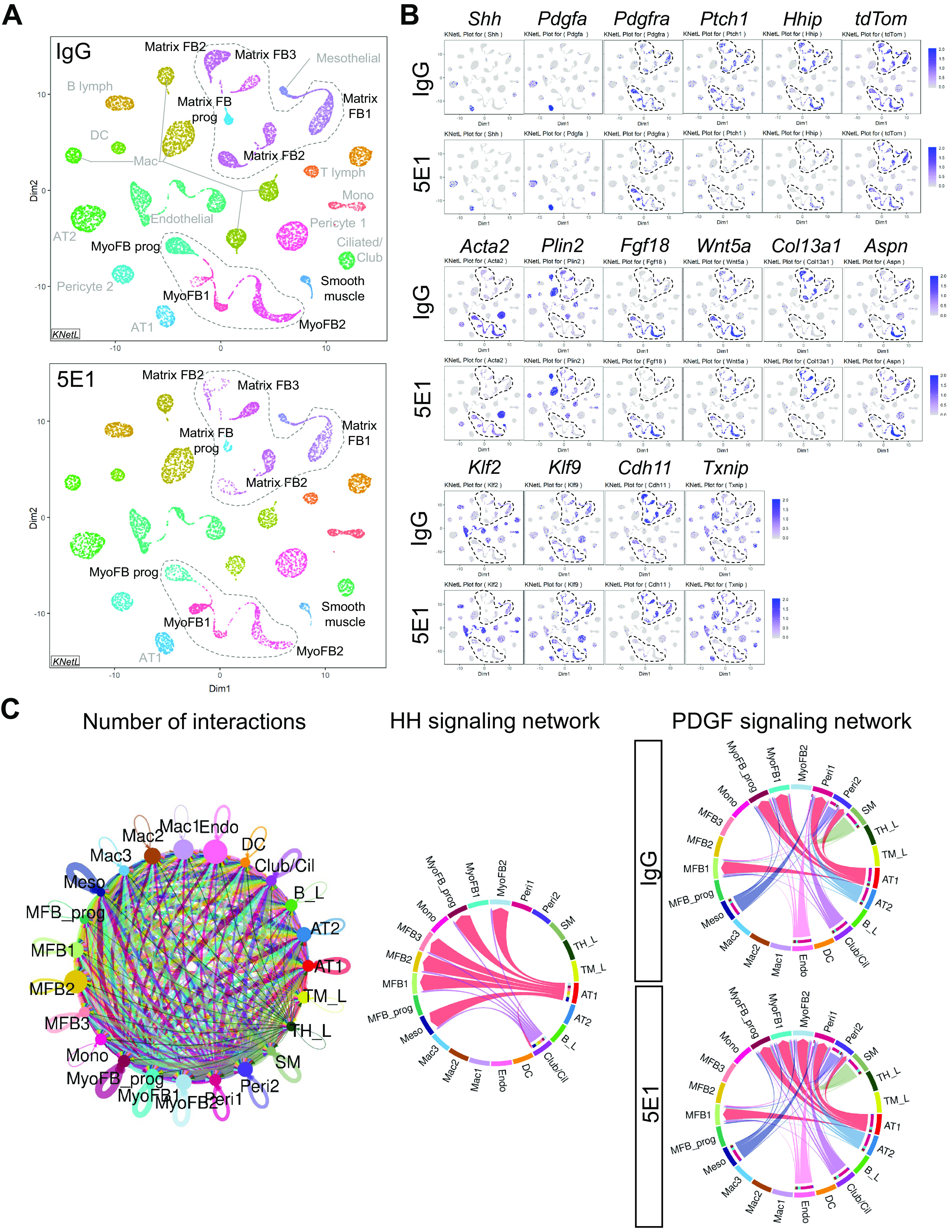
Figure 6.

Single-cell transcriptomic analysis identifies HH–PDGF targets in several postnatal lung mesenchymal cell populations. Whole-lung single-cell suspensions of Gli1creERT2;R26tdTom lungs, lineage labeled at P1 and 5E1 or IgG treated at P3 (n = 3/group), were pooled per condition and loaded onto the 10x Genomics Chromium platform for construction of sequencing libraries. Data analysis was performed using the iCellR pipeline. (A) All major lung cell types were identified by hierarchical clustering, dimensionality reduction, and determination of cluster cell types on the basis of key cell identification markers following the annotations from the LungGENS study (34). Shown are KNetL plots of the 23 clusters, obtained through hierarchical clustering comparing IgG control-treated (top panel) and 5E1-treated (bottom panel) lungs. (B) Selected KNetL plots of 5E1- and IgG-treated lungs illustrate overlap of key pathway genes of HH and PDGF signaling in mesenchymal cell clusters (dotted circles), such as myofibroblasts (MyoFBs) (Acta2) and lipofibroblast (Plin2) and matrix fibroblast (MFB) clusters, and effects of HH inhibition in these clusters. tdTom expression reveals tamoxifen-labeled Gli1 cell lineage. Pdgfra-expressing clusters show changes in PDGF signaling targets (Klf2, Klf9, Txnip, and Cdh11). Gene combinations identify specific clusters; for example, Aspn and Hhip present, Pdgfra absent marks cluster MyoFB2 (potential HH single-receiver cells). (C) Sets of differentially expressed genes for each cluster and condition (5E1 and IgG) were analyzed for ligand receptor interactions using CellChat analysis (see Methods). Chord plots depict the total number of projected interactions between different cells types (left), the cell-specific interactions for the HH signaling network (middle), and the PDGF signaling network with and without HH inhibition (right) with significant communication probability (P < 0.05). Identified are mesenchymal cell types that receive single input of either HH ligand (MyoFB2, MFB2, MFB3, and Meso) or PDGFa ligand (MyoFB1) or combined input from HH and PDGFa (MyoFBprog and MFB1). Of note, cluster MyoFB2 gains PDGFa input with HH inhibition. B_L =B-Lymphocyte; Cil = ciliated; DC = dendritic cell; Endo = endothelial; FB = fibroblast; Mac = macrophage; Meso = mesothelial; Mono = monocyte; Peri = pericyte; SM = smooth muscle; TH_L = T helper lymphocyte; TM_L = T IgM-Fc lymphocyte.
