Figure 2.

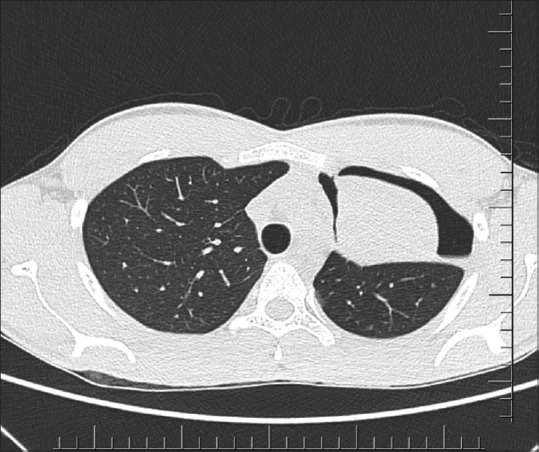
Axial CT thorax image of the second patient showing left upper lobe collapse and shallow pneumothorax surrounding the collapsed lung

Axial CT thorax image of the second patient showing left upper lobe collapse and shallow pneumothorax surrounding the collapsed lung