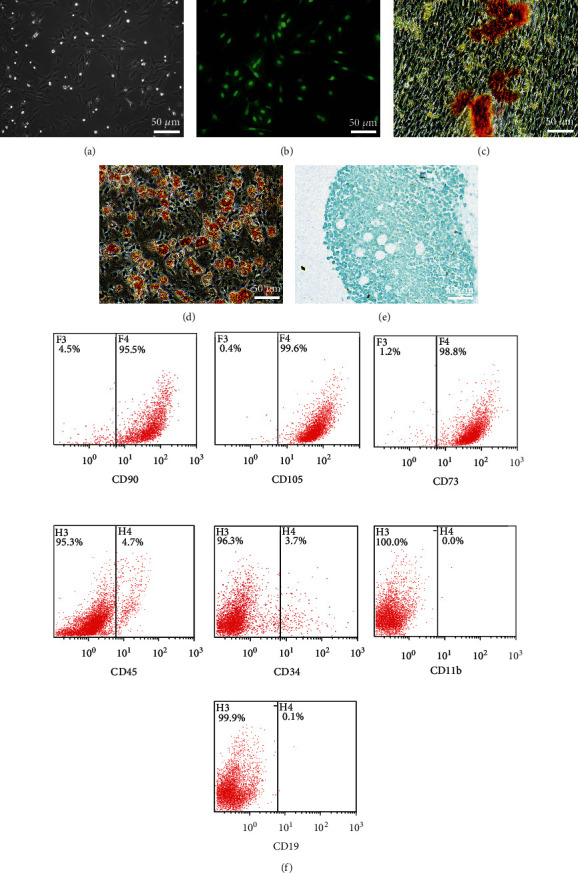
Figure 1.

The characterization of BMSCs. (a) BMSCs were observed using a microscope. BMSC-GFP cells showed green fluorescence (b) under a fluorescence microscope. (c–e) The result of BMSCs cultured in osteogenic, lipogenic, and chondrogenic media for 3 weeks. (f) The rates of CD90, CD105, CD73, CD45, CD34, CD11b, and CD19 positivity on P3 BMSCs.
